Knee
oteotomy is surgery that removes a part of the bone of
the joint of either the bottom of the femur (upper leg
bone) or the top of the tibia (lower leg bone) to increase
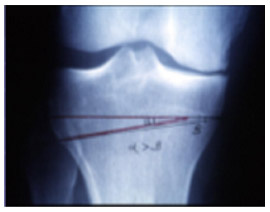
the stability of the knee. Osteotomy redistributes the
weight-bearing force on the knee by cutting a wedge of
bone away to reposition the knee. The angle of deformity
in the knee dictates whether the surgery is to correct
a knee that angles inward, known as a varus procedure,
or one that angles outward, called a valgus procedure.
Varus osteotomy involves the medial (inner) section of
the knee at the top of the tibia. Valgus osteotomy involves
the lateral (outer) compartment of the knee by shaping
the bottom of the femur.
|
|
| |
|
Osteotomy
surgery changes the alignment of the knee so that the weight-bearing
part of the knee is shifted off diseased or deformed cartilage
to healthier tissue in order to relieve pain and increase
knee stability. Osteotomy is effective for patients with
arthritis in one compartment of the knee. The medial compartment
is on the inner side of the knee. The lateral compartment
is on the outer side of the knee. The primary uses of osteotomy
occur as treatment for: |
|
| |
|
-
Knee
deformities such as bowleg in which the knee is varus-leaning
(high tibia osteotomy, or HTO) and knock-knee (tibial
valgus osteotomy), in which the knee is valgus leaning.
-
Osteoarthritis
that includes loss of range of motion, stiffness, and
roughness of the articular cartilage in the knee joint
secondary to the wear and tear of motion, especially
in athletes, as well as cartilage breakdown resulting
from traumatic injuries to the knee. Surgery for progressive
osteoarthritis or injury-induced arthritis is often
used to stave off total joint replacement.
|
|
After
surgery, patients are placed in a hinged brace. Toe-touching
is the only weight-bearing activity allowed for four weeks
in order to allow the osteotomy to hold its place. Continuous
passive motion is begun immediately after surgery and physical
therapy is used to establish full range of motion, muscle
strengthening, and gait training. After four weeks, patients
can begin weight-bearing movement. The brace is worn for
eight weeks or until the surgery site is healed and stable.
X rays are performed at intervals of two weeks and eight
weeks after surgery. |
|
 |
|
|